Multicomponent flow in kidney disease with multi-b-value DWI: GitHub
Purpose: Examine the theory and potential clinical translation of multi-b-value DWI for multicomponent flow in kidney allografts and native kidneys with renal cell carcinoma.
Methods: Multi-b-value DWI (nine b-values; 0–800s/mm2) from a kidney cortex is simulated with anisotropic and non-Gaussian (i.e., anomalous) vas- cular, tubular, and tissue components and analyzed with a Bayesian biexpo- nential, least-squares triexponential, and spectral diffusion MRI. Comparison and application of biexponential, triexponential, and spectral diffusion flow proxies as the product of signal fraction f and diffusion coefficient D, fD for each component, is demonstrated in studies of kidney disease and compared to clinical measures.
Results: Simulated spectral diffusion fD demonstrated strong correlation to input fD of the simulated anisotropic and anomalous components. It agreed with both three-component diffusion and two-component diffusion. fD showed similar or improved agreement and correlation to input than the individual parameters f and D, and spectral diffusion showed similar or improved agreement than corresponding bi- and triexponential models. n kidney allografts, fD from spectral diffusion showed that allografts with higher fibrosis score had higher fDtissue while allografts with impaired function had reduced fDtubule. IVIM perfusion fraction f and fD* correlated with helper T-cells and T-cells as well as macrophages in renal clear cell carcinoma while kidney DWI predicted decline in function after nephrectomy of renal masses.
Conclusions: in kidney allografts, higher fDtissue may capture the collagen and scarred fibrotic tissue by the greater amount of the restricted diffusion. Allografts with impaired function with reduced fDtubule suggests detection of reduced tubular flow and filtration. DWI detected fibrosis in asymptomatic patients suggesting potential detection of earlier microstructural disease.
Multi-b-value DWI also demonstrated promise in immunophenotyping of renal cell carcinoma and pre-operative detection of compensatory flow indicative of poor response to nephrectomy.
Example visualization of simulated curves, multicompartment diffusion maps of a healthy volunteer, and sensitivity to clinical measures.

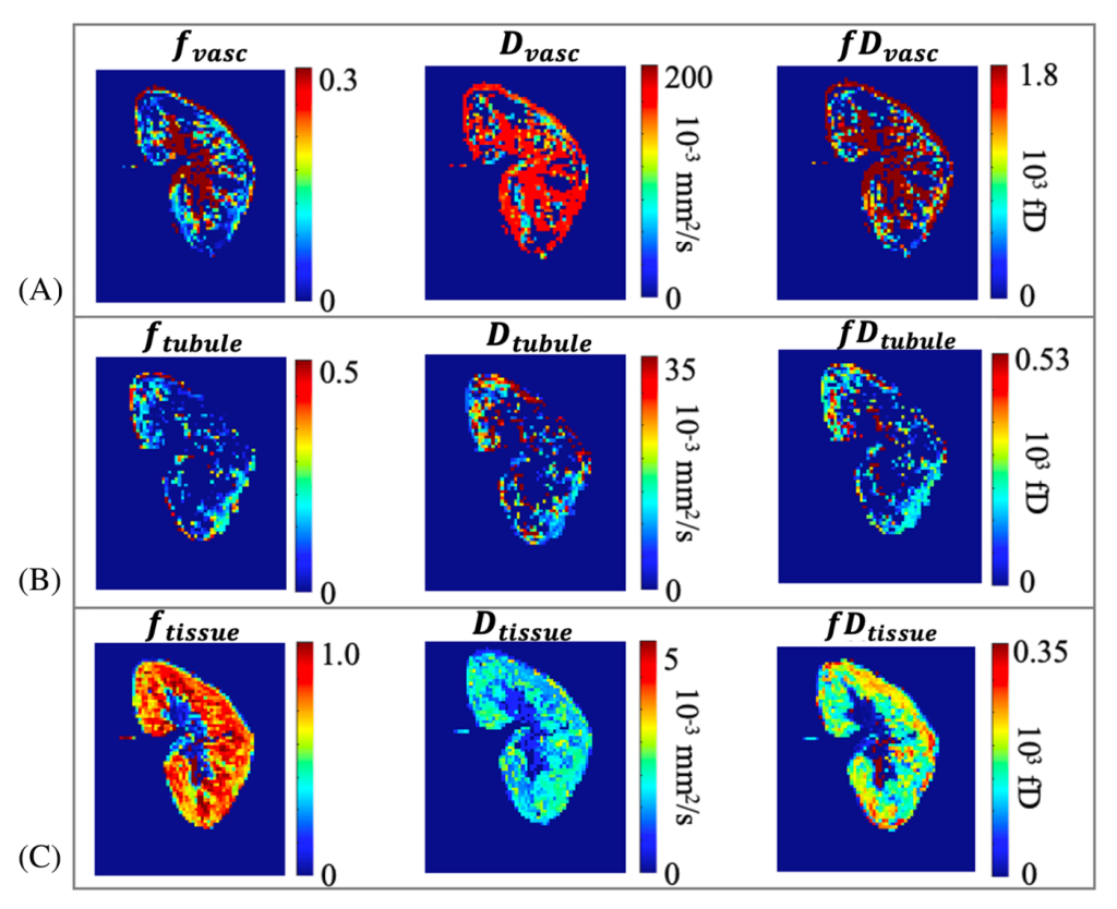
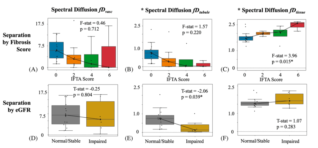
Figures from “Estimation of Multi-Component Flow in the Kidney with Multi-b-value Spectral Diffusion” Magnetic Resonance in Medicine 2025
List of some publications relevant to this topic:
M. Liu, T. Gladytz, J. Dyke et al. Estimation of Multi-Component Flow in the Kidney with Multi-b-value Spectral Diffusion. Magn Reson Imaging. 2025. 10.1002/mrm.30644
M. Liu, J. Dyke, T. Gladytz et al. Detecting early kidney allograft fibrosis with multi-b-value spectral diffusion MRI. Sci Rep. 10.1038/s41598-025-24701-5
JS Periquito, T. Gladytz, JM Millward et al. Continuous diffusion spectrum computation for diffusion-weighted magnetic resonance imaging of the kidney tubule system. Quantitative Imaging in Medicine and Surgery. 2021. 10.21037/qims-20-1360.
J. Stabinska, A. Ljimani, HJ Zöllner et al. Spectral diffusion analysis of kidney intravoxel incoherent motion MRI in healthy volunteers and patients with renal pathologies. Magnetic Resonance in Medicine. 2021. 10.1002/mrm.28631.
M. Liu, O. Bane, X. Mu. et al. Multiparametric MRI for Predicting Renal Function Deterioration and Chronic Kidney Disease Development
in Patients Undergoing Nephrectomy for Renal Masses:
A Pilot Study, 2025. DOI: 10.1002/jmri.70213
M. Liu, O. Bane, X. Mu. et al. Immuno-Oncologic Profiling of Renal Masses using Multiparametric MRI: A Pilot Study. Journal of ImmunoTherapy of Cancer, 2025; DOI: 10.1136/jitc-2025-012833
J. Stabinska, HJ Wittsack, LO Lerman, et al. Probing Renal Microstructure and Function with Advanced Diffusion MRI: Concepts, Applications, Challenges, and Future Directions. Journal of Magnetic Resonance Imaging. 2023;doi:10.1002/jmri.29127
EE Sigmund, PH Vivier, D Sui, et al. Intravoxel Incoherent Motion and Diffusion-Tensor Imaging in Renal Tissue under Hydration and Furosemide Flow Challenges. Radiology. 2012. 10.1148/radiol.12111327.
A. Ljimani, A. Caroli, C. Laustsen et al. Consensus-based technical recommendations for clinical translation of renal diffusion-weighted MRI. Magnetic Resonance Materials in Physics, Biology and Medicine. 2019. 10.1007/s10334-019-00790-y.
A. Caroli, M. Schneider, I. Friedli, et al. Diffusion-weighted magnetic resonance imaging to assess diffuse renal pathology: a systematic review and statement paper. Nephrology Dialysis Transplantation. 2018. doi:10.1093/ndt/gfy163
O. Bane, M. Wagner, JL Zhang, et al. Assessment of renal function using intravoxel incoherent motion diffusion‐weighted imaging and dynamic contrast‐enhanced MRI. Journal of Magnetic Resonance Imaging. 2016; doi:10.1002/jmri.25171
R. van der Bel, OJ. Gurney-Champion, M. Froeling et al. A tri-exponential model for intravoxel incoherent motion analysis of the human kidney: In silico and during pharmacological renal perfusion modulation. European Journal of Radiology. 2017. doi:10.1016/j.ejrad.2017.03.008
Federau C. Intravoxel incoherent motion MRI as a means to measure in vivo perfusion: A review of the evidence. NMR in Biomedicine. 2017; doi:10.1002/nbm.3780
AL Liu, A Mikheev, H. Rusinek H, et al. REnal Flow and Microstructure AnisotroPy (REFMAP) MRI in Normal and Peritumoral Renal Tissue. Journal of Magnetic Resonance Imaging. 2018. doi:10.1002/jmri.25940
M. Liu, O. Bane, H. Al-Mubarak, A. Reddy, P. Kennedy, P. Robson, J. Cuevas, K. Meilika, A. Horowitz, B. Kuhn, K. Badani, B. Taouli, S. Lewis. Assessment & Prediction of Renal Function with Non-Contrast MRI in Patients Undergoing Surgical Management of Solid Renal Masses.” International Society for Magnetic Resonance in Medicine Workshop on IVIM 2024. (Oral Presentation)
